Helpline
8420008000
Donate Your Eyes
COVID-19 Protocol

- About Us
- Medical Services
- Hospital Facilities
- Patient Info
- Know your Eyes
- News & Events
- People
- Contact


- Home
- /
- Know your Eyes
- /
- Anatomy of the Eye
Anatomy of the Eye
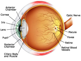
The eye is a complex organ composed of many small parts, each vital to normal vision. The ability to see clearly depends on how well these parts work together. It is actually an extension of the brain, connected by the optic nerve, which consists of thousands of nerve fibers. These nerves carry visual messages, in the form of nerve impulses, from the retina to the brain, which interprets the impulses as images.
Layers of the Eye
There are three layers of tissue in the walls of the eye. They are:

The Outer Layer - Sclera & Cornea
The Sclera
The sclera, or white visible portion of the eye, forms the outermost layer of tissue of the posterior and lateral aspects of the eyeball. It consists of a firm fibrous membrane that maintains the shape of the eye and attachment to the extraocular or extrinsic muscles of the eye.
The Cornea
The clear, dome-shaped surface that covers the front of the eye. Being the front window of the eye, it is the main source of refraction. The cornea has the dual purpose - protecting the eye and refracting light as it enters the eye. The cornea is as strong and durable as plastic and as smooth and clear as glass, which means it is also an excellent shield against dust, germs and other debris.
While the cornea contains no blood vessels, it is packed with nerve fibers, making it highly sensitive to pain.
The Middle Vascular Layer - Choroid, Ciliary body & Iris
The Choroid
The thin blood-rich membrane, that lies between the retina and the sclera. It is made up of blood vessels that provide nourishment to the eye and is responsible for supplying blood to the retina.
The Ciliary Body
Is a part of the eye that produces aqueous humor. It contains two main structures - the first is a muscle that contracts and expands to control the curvature of the lens during accommodation and the second is a gland that secretes aqueous humor.
The Iris
The colored part of the eye. It is partly responsible for regulating the amount of light permitted to enter the eye. The iris has three layers and is distinctive because of its genetically determined coloring.
The Inner Nervous Tissue Layer - Retina
The Retina
This membrane lines the inside wall of the eye. It contains photoreceptors (rods and cones) that change light into sight by converting light into electrical impulses. These electrical messages are sent from the retina to the brain and are interpreted as images.
Other parts
THE LENS
Immediately behind the iris is the Lens, it is the transparent structure inside the eye that focuses light rays onto the retina. Located just behind the pupil it allows for changing of focus from distance to near objects by altering its shape. This changing focus is called Accommodation. As people age, the lens can harden, decreasing focusing ability.
THE PUPIL
The opening in the middle of the iris through which light passes to the back of the eye. It is the light sensitive nerve layer that lines the back of the eye. Its size changes since its function is to control the amount of light reaching the retina. In the dark, it expands allowing more light to enter. It contracts in bright light to keep out excess light.
THE OPTIC NERVE
A bundle of nerve fibers that connect the retina with the brain. The optic nerve carries signals of light, dark, and colors to the area of the brain (the visual cortex), which assembles the signals into images. This nerve is the pathway that the light rays take from the retina to the processing centre of the brain. It actually is made of about a million tiny nerves bundled together.
THE OPTIC DISC
This area is not sensitive to light and it is often referred to as the "blind spot". It is where the retina meets the optic nerve.
THE ANTERIOR CHAMBER
The front section of the eye's interior where aqueous humor flows in and out of providing nourishment to the eye and surrounding tissues.
THE AQUEOUS HUMOR
Is a clear watery fluid in front of the eyeball. This fluid is produced by the ciliary body and circulates in the front part of the eye. It provides nourishment to the front parts of the eye and maintains the eye pressure.
THE BLOOD VESSELS
TUBES
Arteries and veins that carry blood to and from the eye.
THE CARUNCLE
A small, red portion of the corner of the eye that contains modified sebaceous and sweat glands.
UPPER EYELID
Top, movable, superior fold of skin that covers the front of the eyeball when closed, including the cornea.
LOWER EYELID
Lower, inferior, skin that covers the front of the eyeball when closed.
THE MACULA
This tiny part of the retina is the central focusing spot. It allows us to see minute details clearly and is also responsible for color vision.
POSTERIOR CHAMBER
The back part of the eye's interior.
SUSPENSORY LIGAMENT OF LENS
A series of fibers that connect the ciliary body of the eye with the lens, holding it in place.
THE VITREOUS BODY
A clear, jelly-like, substance that fills the back part of the eye. This clear gel fills the central core of the eye. It helps to maintain a spherical shape to the eye.


